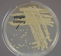
Results: streak plate method and pure culturesCS11-15.jpeg
Dr. Spencer: Well-done, you have isolated pure, single colonies! Now that we've isolated single colonies, what's our next step in studying these to identify the causative pathogen(s)? Sam: First, we should observe the colonies themselves. We need to pay attention to characteristics like shape, size, color, and texture. These morphological traits can provide us with initial identification clues. You: Absolutely, Sam. Observing the colony morphology may help us differentiate various bacterial types, even before performing other tests. Dr. Spencer: Good. After colony morphology, what about their cellular characteristics? Sam: Next, we move to microscopy. We'll prepare either wet mounts or fixed smears to examine our bacterial cells under the microscope. This will allow us to see if they are rod-shaped or spherical. You: We could then proceed with Gram staining the samples. It's a quick and effective way to differentiate bacteria into Gram-positive and Gram-negative categories. Sam: Right, and that classification gives us a solid direction on narrowing down our potential pathogen candidates. Selective and differential media can also be used to further our study. You: Precisely, and finally, after identification, conducting antibiotic susceptibility tests can determine the best treatment approach for the patient. Dr. Spencer: Excellent summary. In this way, we can identify the pathogen(s) and tailor effective interventions. |
Map: CS11 - MICROBIAL CULTURE AND GROWTH _ES (1054)
|
||
|
Review your pathway |