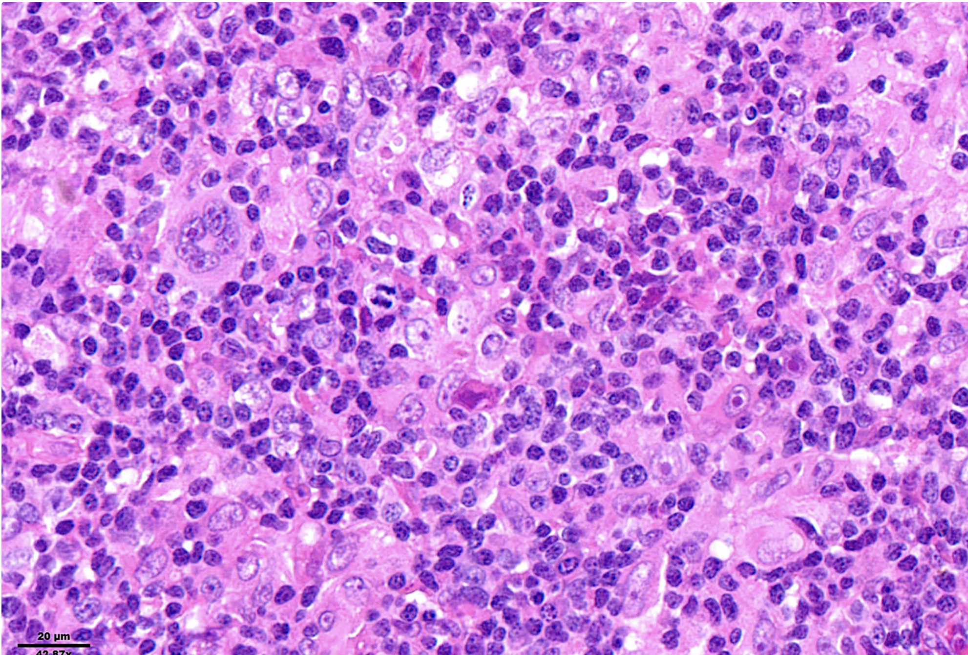
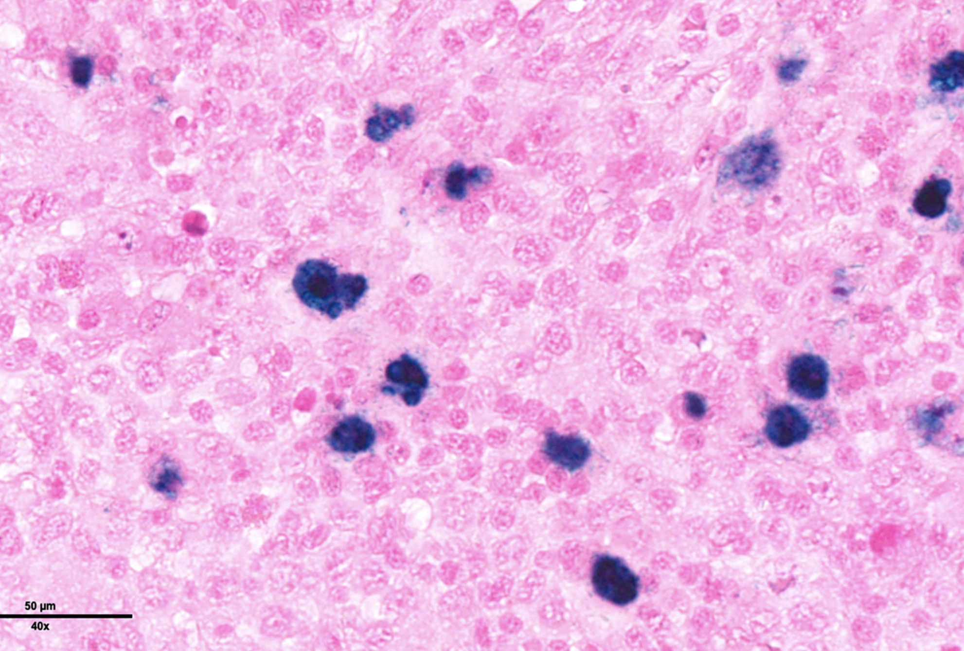
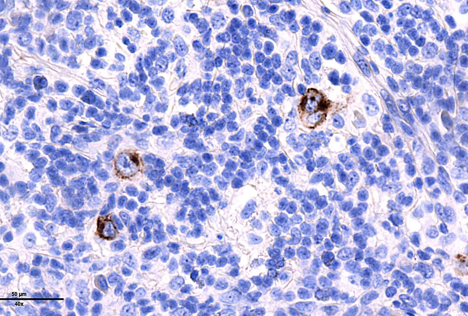
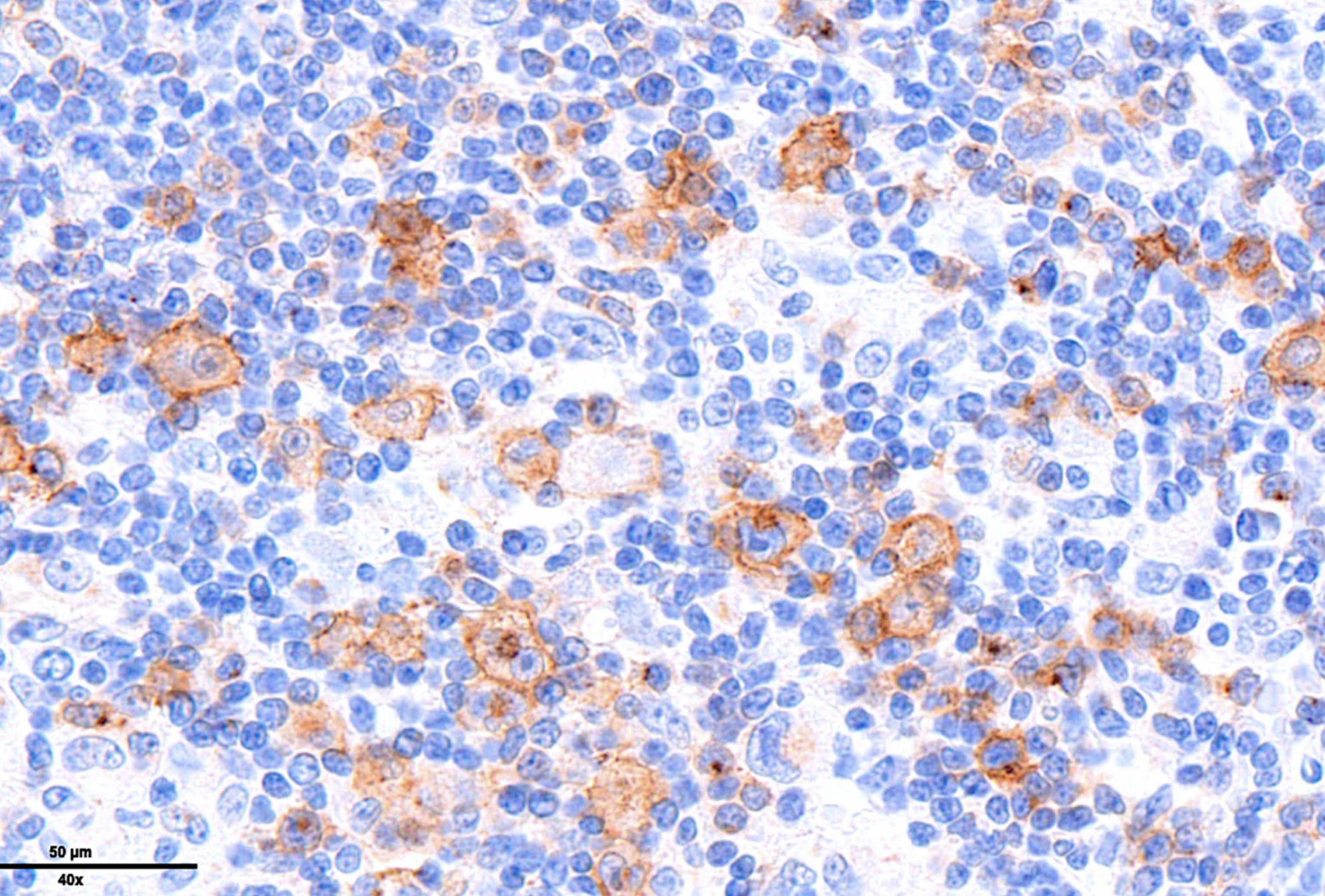
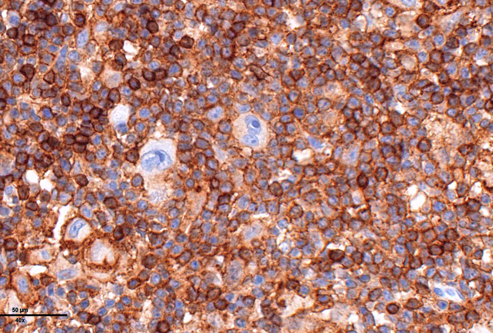
Lymph Nodes sample Mixed cellularity classic Hodgkin lymphoma. H&E stained sections show a lymph node with architectural effacement by an interfollicular mixed inflammatory infiltrate comprising small lymphocytes, eosinophils, neutrophils, plasma cells, with scattered admixed large atypical lymphoid cells. The neoplastic cells have monolobated / bilobated / multilobated nuclei, vesicular chromatin, prominent nucleoli and amphophilic cytoplasm, consistent with Hodgkin and Reed-Sternberg cells. Immunophenotypically, the large atypical cells are positive for PAX5 (weak), CD30 (strong), negative CD20 and CD45. ( CD30 fig.4/ CD45 fig.5) In situ hybridization for EBV encoded RNA (EBER) is positive on the large atypical cells. (fig.2/ 3 EBV-LMP1) |
Map: LLA in 8 years old Child (1046)
|
||
|
Review your pathway |